
Medical imaging generates vast volumes of complex visual data every day. Radiologists examine hundreds of scans during a typical shift, searching for microscopic subtle structural abnormalities that signal early stage disease.
Computer Aided Detection (CADe) serves as an automated technological safety net. By analyzing digital scans alongside human physicians, these systems highlight suspicious regions to reduce oversight errors, speed up workflow evaluation, and catch subtle anomalies before clinical symptoms appear.
What Is Computer Aided Detection?
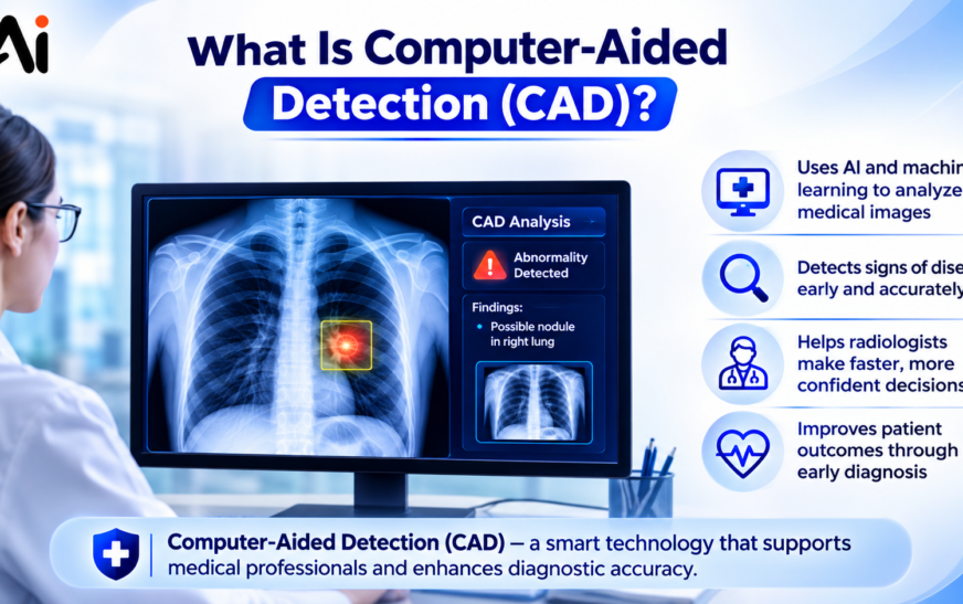
Computer Aided Detection refers to specialized software algorithms designed to analyze medical images and identify potential regions of interest that require human review.
Definition of Computer Aided Detection
Computer Aided Detection is an interdisciplinary technology combining digital image processing, pattern recognition, and artificial intelligence to assist medical professionals.
The primary goal of CADe is to mark suspicious features on diagnostic scans. It does not attempt to diagnose whether a mass is benign or malignant. Instead, it functions as a digital second reader, drawing the radiologist’s attention to areas that exhibit abnormal density, asymmetry, microcalcifications, or structural distortion.
Why Computer Aided Detection Matters
Visual fatigue and cognitive overload represent real challenges in diagnostic radiology. Studies show that between 10% and 30% of early lesions are missed during routine initial screenings due to observational oversights or complex overlapping tissue structures.
Computer Aided Detection addresses these human visual limitations by processing every pixel uniformly. It applies consistent mathematical evaluation standards across all images regardless of case volume or shift duration.
When subtle abnormalities are highlighted early, patients receive timely interventions that significantly improve long term survival rates.
A Brief History of Computer Aided Detection
Early research into automated medical image analysis began in the late 1960s and 1970s at academic research institutions. Early computational limitations restricted these initial systems to basic edge detection and simple thresholding.
Development accelerated rapidly throughout the 1980s with the advent of digital radiography and improved computing architectures.
The U.S. Food and Drug Administration reached a major milestone in 1998 when it approved the first commercial CADe system for screening mammography. Following Medicare reimbursement approvals in 2002, clinical adoption expanded across hospitals and imaging centers worldwide.
How Computer Aided Detection Works
Modern CADe platforms process medical imagery through a multi-stage sequential workflow designed to isolate abnormal anatomical signals from complex anatomical noise.
The overall detection pipeline relies on the following operational steps:
- Capturing high resolution digital data from diagnostic imaging devices.
- Preprocessing images to remove noise and standardize contrast levels.
- Segmenting relevant anatomical structures from surrounding background tissue.
- Extracting textural, geometric, and density feature metrics.
- Applying classification models to identify suspicious patterns.
- Rendering visual markers on the radiologist’s display terminal.
Image Acquisition
The detection pipeline begins when a patient undergoes a diagnostic scan. Raw signals from X-ray detectors, computed tomography sensors, or magnetic resonance coils are converted into digital DICOM files. High spatial resolution and proper signal to noise ratios are critical during this phase because low quality source data degrades downstream analytical performance.
Image Processing and Analysis
Once DICOM files enter the CADe server, automated preprocessing routines clean the image. Software algorithms apply equalization filters to harmonize contrast variations across different exposure settings. Advanced segmentation routines then isolate target organs, such as separating lung parenchymal tissue from chest wall bone structures.
Pattern Recognition and Abnormality Detection
Isolating suspicious features relies on complex pattern recognition mathematics. The algorithm measures localized parameters including shape compactness, edge sharpness, gray level gradients, and spatial symmetry relative to adjacent tissue.
When evaluating calcifications, the system searches for tiny clusters of high density pixels measuring less than 1 millimeter in diameter. When evaluating soft tissue, it screens for irregular mass borders, focal architectural distortions, or central density spikes.
Highlighting Suspicious Areas for Review
If a detected region crosses predefined mathematical probability thresholds, the system places visual markers over the image. These marks appear as geometric outlines, arrows, or bounding boxes on the radiologist’s workstation screen.
The radiologist reviews these flagged locations alongside the original unannotated scan before making a final clinical interpretation.
The Role of Artificial Intelligence and Machine Learning
Early CADe applications used hand crafted rules written by software engineers. Modern platforms utilize advanced machine learning models trained on millions of annotated clinical cases.
Convolutional neural networks extract thousands of subtle structural features automatically without requiring manual feature programming. This transition to deep learning has improved detection sensitivity while gradually reducing background false positive rates.
Key Components of a Computer Aided Detection System
A complete CADe deployment relies on four interconnected operational layers working together in real time.
The necessary structural components include:
- Diagnostic hardware devices that generate raw digital patient images.
- High speed computing servers running image processing software pipelines.
- Machine learning model engines that run pattern classification algorithms.
- Interactive physician review workstations connected to Picture Archiving and Communication Systems.
Medical Imaging Devices
The primary source hardware includes digital mammography units, computed tomography scanners, magnetic resonance imaging machines, digital X-ray detectors, and ultrasound transducers. These systems generate high precision pixel grids that feed raw data directly to analytical processing nodes.
Image Processing Software
The software layer manages data transfer protocols, image normalization routines, and structural segmentation routines. It ensures that scans coming from different hardware manufacturers meet uniform formatting standards before algorithmic feature extraction begins.
AI and Machine Learning Algorithms
The computational engine executes statistical classification models. It compares regional image vectors against learned statistical patterns to differentiate normal anatomical variance from true pathological abnormalities.
Radiologist or Healthcare Professional Review
The human specialist remains the final decision maker. CADe outputs serve purely as diagnostic aids. The treating clinician correlates highlighted marks with patient medical history, physical examination findings, and previous historical imaging prior to signing the diagnostic report.
Types of Computer Aided Detection
CADe systems are customized to analyze specific anatomical regions and targeted disease processes.
Mammography Computer Aided Detection
Screening mammography represents the most widely deployed CADe application globally. Algorithms screen two dimensional digital mammograms and three dimensional digital breast tomosynthesis slices.
These platforms achieve sensitivity rates up to 99% for detecting microcalcification clusters and between 75% and 89% for identifying soft tissue masses.
Lung Cancer Detection
Thoracic CADe applications analyze chest radiography and low dose computed tomography scans. Software searches for solitary pulmonary nodules ranging from 3 millimeters to 30 millimeters in diameter. Early identification of small ground glass nodules allows oncologists to diagnose stage I lung carcinomas when surgical cure rates remain high.
Colon Cancer Detection
Virtual colonoscopy systems utilize CADe software to inspect three dimensional reconstruction models from CT colonography scans. Algorithms screen the mucosal surface of the colon to detect flat and pedunculated adenomatous polyps. Automated detection of pre-cancerous polyps larger than 6 millimeters significantly reduces invasive screening burdens for patients.
Brain Imaging Analysis
Neuroimaging CADe modules analyze magnetic resonance imaging and computed tomography scans of the central nervous system. Systems assist in identifying hyperdense ischemic stroke regions, cerebral microbleeds, intracranial aneurysms, and localized cortical atrophy patterns associated with neurodegenerative conditions.
Prostate Cancer Detection
Multi-parametric MRI analysis relies on CADe algorithms to evaluate prostate tissue architecture. The software evaluates T2 weighted sequences, diffusion weighted imaging, and dynamic contrast enhanced scans simultaneously. Flagging suspicious regions helps urologists target magnetic resonance guided biopsies with millimeter accuracy.
Dental Imaging Detection
Dental CADe tools analyze panoramic X-rays and cone beam computed tomography scans. These systems automatically highlight interproximal caries, subtle periapical bone loss, impacted root structures, and periodontal bone absorption lines during routine examinations.
Benefits of Computer Aided Detection
Integrating CADe tools into routine clinical workflows yields clear diagnostic and operational advantages for healthcare organizations.
Key operational benefits include:
- Detecting microscopic disease indicators before visible physical symptoms emerge.
- Increasing overall diagnostic sensitivity across broad patient screening populations.
- Streamlining image interpretation workflows for busy radiological departments.
- Minimizing missed findings caused by human visual fatigue or distraction.
- Improving long term clinical survival metrics through early therapeutic intervention.
Earlier Disease Detection
Screening programs aim to detect illness at its earliest stage. CADe algorithms pick up subtle architectural distortions and minor density shifts that can escape initial visual inspection. Catching malignancies early reduces treatment intensity and improves patient recovery outlooks.
Improved Diagnostic Accuracy
Adding a digital second reader elevates diagnostic sensitivity across clinical environments. Junior radiologists and general practitioners benefit from computational prompts that mirror the observational consistency of specialized subspecialist readers.
Increased Efficiency for Healthcare Professionals
Radiologists must read large volumes of complex cross sectional imaging daily. CADe software auto-segments normal tissue structures and directs clinician attention directly to high risk image slices, saving valuable interpretation time per case.
Reduced Human Error
Human visual attention fluctuates during long work shifts. CADe systems maintain absolute observational consistency, processing case number one and case number one hundred with identical mathematical precision to catch oversights.
Better Patient Outcomes
Earlier detection correlates directly with improved survival statistics. When screening mammography, lung CTs, and colon scans identify pre-invasive lesions promptly, patient curative success rates rise substantially.
Limitations of Computer Aided Detection
Automated detection tools are not flawless. Every system operates within algorithmic limits established by training data, pixel resolution, and optical image noise.
False Positives
A false positive occurs when the software incorrectly marks healthy tissue or benign structures as suspicious.
In screening mammography, standard CADe platforms can generate between 0.5 and 2.0 false positive prompts per image frame.
These extra marks require radiologists to spend additional time evaluating non-pathological areas. High false positive counts can increase physician fatigue, lead to unnecessary patient recall appointments, and cause unneeded emotional distress.
False Negatives
A false negative occurs when the software fails to mark a genuine pathological lesion.
Dense breast tissue, subtle ground glass pulmonary opacity, and irregular anatomical shadows can obscure abnormal features from algorithmic filters.
Radiologists who rely too heavily on automated prompts risk overlooking lesions that the software missed. Maintaining independent primary review protocols remains vital to prevent false negative clinical oversights.
Dependence on Image Quality
Algorithmic performance relies directly on source image quality.
Low spatial resolution, patient motion artifacts, incorrect radiation exposure settings, and improper patient positioning degrade detection precision.
If raw digital image input is noisy or blurry, feature extraction filters cannot differentiate subtle microcalcifications from background sensor artifacts.
Not a Replacement for Medical Experts
Computer aided detection tools possess no genuine clinical understanding. They process statistical pixel variations without understanding patient medical history, genetic risk markers, or physical symptoms.
The software acts purely as an observational pointer. Medical diagnosis requires human clinical reasoning, contextual analysis, and comprehensive patient evaluation.
Cost and Implementation Challenges
Deploying enterprise detection software requires substantial financial investment.
Hospital expenditures associated with deployment include the following items:
- Purchasing commercial software licenses and server infrastructure.
- Upgrading local Picture Archiving and Communication Systems storage servers.
- Training radiology staff and IT engineers on new software tools.
- Paying ongoing software maintenance and algorithm update fees.
Computer Aided Detection vs. Computer Aided Diagnosis
In medical imaging literature, technical descriptions divide automated image processing platforms into two distinct operational classes known as CADe and CADx.
Key Differences
Computer Aided Detection, abbreviated as CADe, focuses exclusively on localization. It identifies where a potential abnormality sits on a scan and marks it for human inspection.
Computer Aided Diagnosis, abbreviated as CADx, goes a step further by evaluating what the lesion actually is. It calculates probability scores regarding disease severity, such as estimating whether a lung nodule is benign or malignant.
CADe answers the location question, while CADx answers the characterization question.
When Each System Is Used
Healthcare facilities use CADe during primary population screening programs where high volumes of asymptomatic patients require quick visual triage. Mammography centers and chest X-ray screening clinics rely heavily on CADe.
Clinicians use CADx in secondary diagnostic workups. When a suspicious mass is already confirmed, CADx software analyzes lesion texture, margins, and contrast uptake to help determine whether a patient needs an invasive tissue biopsy.
Which One Is More Effective?
Neither system replaces the other because both address separate stages of the diagnostic pipeline.
CADe excels at finding hidden needle in a haystack lesions across thousands of routine images. CADx excels at offering objective quantitative measurements to support complex treatment decisions.
Combining both platforms into unified multi-stage software suites offers the highest overall diagnostic support.
Real-World Applications of Computer Aided Detection
Clinical utilization of CADe spans multiple major medical specialties.
Breast Cancer Screening
Breast imaging represents the largest clinical market for detection software, accounting for over 66% of global CADe application revenue.
Systems analyze digital mammograms and 3D tomosynthesis volumes to highlight clusters of microcalcifications, soft tissue masses, and architectural distortions. Integrated detection software improves early invasive cancer retrieval by 8% to 15% in population screening cohorts.
Lung Disease Detection
Thoracic CADe software analyzes chest X-rays and low-dose computed tomography scans.
It identifies early pulmonary nodules, focal consolidations, ground-glass opacities, and signs of active tuberculosis. In global public health initiatives, automated chest X-ray detection systems help triage high volumes of respiratory cases efficiently.
Cardiovascular Imaging
Cardiovascular detection tools process computed tomography angiography scans to identify coronary artery calcification levels.
Software algorithms highlight luminal narrowing, calculate total coronary calcium scores, and detect subtle aortic dissection lines before critical vascular events occur.
Neurology and Brain Disorders
Neuroimaging platforms assist emergency radiologists by flagging acute intracranial hemorrhages, ischemic stroke arterial blockages, and cerebral aneurysms on non-contrast brain CT scans.
Rapid automated alerting speeds up emergency thrombolytic treatments, preserving brain tissue during critical medical windows.
Dental Diagnostics
Modern dental clinics use automated image analysis to review digital panoramic X-rays.
The algorithms automatically flag interproximal tooth decay, periapical abscess formations, impacted wisdom teeth, and early alveolar bone degradation, ensuring consistent routine evaluations.
Orthopedic Imaging
Orthopedic detection tools analyze musculoskeletal radiographs to identify subtle non-displaced fractures, joint space narrowing, bone mineral density shifts, and loosening around prosthetic implants.
The Role of Artificial Intelligence in Computer Aided Detection
Artificial intelligence has transformed medical imaging from rigid rule-based programming into dynamic, self-improving pattern detection.
How AI Improves Detection Accuracy
Traditional CADe tools relied on hand-coded mathematical rules defined by software developers. These early systems struggled with normal anatomical variations, producing high false positive rates.
Modern AI models learn complex structural relationships directly from massive medical image datasets. They adjust internal feature weights dynamically, resulting in lower false positive rates and higher sensitivity across diverse patient populations.
Deep Learning in Medical Imaging
Deep learning architectures, particularly multi-layer Convolutional Neural Networks (CNNs) and Vision Transformers, process images at multiple abstraction levels.
The lower network layers detect simple pixel boundaries, edges, and density transitions. The deeper network layers synthesize these basic edges into complex three-dimensional anatomical models, allowing the system to recognize faint lesions hidden behind overlapping bone or dense organ tissue.
Future of AI-Powered Detection Systems
Next generation detection models incorporate multi-modal data streams. Instead of analyzing a single image in isolation, upcoming algorithms evaluate current scans alongside historical imaging, genetic risk profiles, and laboratory blood results to calculate holistic patient risk markers in real time.
Industries Using Computer Aided Detection Beyond Healthcare
The core mathematical principles behind CADe, such as digital signal processing, spatial segmentation, and automated anomaly classification, apply widely across non-medical industries.
Manufacturing Quality Inspection
Industrial plants use optical and X-ray CADe systems to check manufacturing quality on high-speed assembly lines.
Automated vision systems inspect semiconductor microchips, printed circuit boards, and metallic castings for microscopic surface cracks, solder bridges, or internal voids without stopping production.
Security and Surveillance
Transport hubs deploy automated threat detection software on baggage X-ray scanners and millimeter-wave body scanners.
The software automatically flags weapons, explosives, organic contraband, and dangerous goods passing through airport screening lanes, assisting security personnel in real time.
Aerospace and Aviation
Aerospace engineers use non-destructive testing CADe software to inspect turbine blades, airframe composite panels, and jet engine welds.
High-resolution industrial CT scans are processed automatically to detect microscopic fatigue cracks and internal delamination before components are installed on aircraft.
Automotive Industry
Autonomous vehicles use vision-based detection systems that operate on the same pattern recognition principles as medical CADe.
Onboard cameras and LiDAR sensors process spatial data continuously to detect pedestrians, lane markers, road signs, and sudden obstacles in real time.
Scientific Research
Astronomers and environmental scientists use automated image detection to analyze vast spatial datasets.
In astronomy, algorithms scan deep-sky telescope images to discover distant exoplanets, asteroids, and supernovae. In climate science, satellite imaging platforms automatically detect deforestation patterns, glacial retreat rates, and wildfire outbreaks.
Common Challenges in Implementing Computer Aided Detection
Integrating automated detection tools into existing hospital operational workflows involves several practical challenges.
Data Privacy and Security
Healthcare imaging datasets contain sensitive protected health information. Transferring DICOM images to cloud-based CADe servers requires strong end-to-end encryption, strict access permissions, and full compliance with regulations like HIPAA in the United States and GDPR in Europe.
Regulatory Compliance
Before entering clinical practice, medical CADe tools must receive formal clearance from regulatory agencies such as the U.S. Food and Drug Administration or obtain a CE mark in Europe.
Developers must present extensive clinical trial data proving their algorithm operates safely and effectively across different hardware platforms and patient demographics.
Integration with Existing Healthcare Systems
Hospital IT networks rely on complex PACS, RIS, and Electronic Health Record architectures.
Detection software must integrate smoothly with these existing systems without causing network latency, crashing diagnostic workstations, or requiring radiologists to switch between separate software interfaces.
Training and Adoption
Introducing new software tools requires comprehensive staff training.
Radiologists must learn how to interpret algorithmic markers correctly without developing over-reliance or automation bias, where a reader uncritically accepts software outputs without independent review.
Best Practices for Using Computer Aided Detection
Following established clinical protocols ensures that CADe tools improve diagnostic accuracy without compromising safety.
Recommended operational guidelines include the following steps:
- Conducting an independent primary review of all unannotated images before turning on CADe overlays.
- Enforcing strict image quality control standards across all diagnostic hardware.
- Keeping software models updated to incorporate the latest algorithm improvements.
- Tracking system false positive and false negative metrics through regular internal audits.
Combine CAD with Expert Review
The most effective workflow uses CADe as a true second reader. The radiologist performs an initial evaluation of the unannotated scan, forms an independent impression, and then toggles on the CADe markers to check for missed areas before signing the final report.
Use High-Quality Medical Images
High-performance detection models require high-quality input data.
Hospital imaging facilities must perform routine calibration on X-ray detectors, CT scanners, and MRI coils to ensure sharp contrast, low noise, and proper exposure parameters.
Regularly Update AI Models
Medical software developers release periodic updates that refine algorithms, reduce false positive rates, and expand detection capabilities. Hospital IT departments should apply validated software patches promptly to keep performance high.
Monitor System Performance
Clinical directors should track internal CADe metrics over time. Comparing software flags against final pathology biopsy results helps hospitals identify potential algorithm drift or hardware calibration issues early.
Future Trends in Computer Aided Detection
Technological advancements continue to expand the scope and speed of automated medical image analysis.
AI-Assisted Clinical Decision Support
Future CADe systems will integrate directly into enterprise clinical decision support networks.
Instead of generating simple visual bounding boxes, software will cross-reference highlighted image anomalies with laboratory test results, patient family history, and electronic health records to suggest tailored diagnostic next steps.
Cloud-Based Detection Systems
Cloud computing allows smaller regional clinics and rural hospitals to access advanced detection platforms without buying expensive local server hardware. Scans are transmitted securely to high-speed cloud servers, analyzed in seconds, and returned with clear annotations.
3D and Real-Time Image Analysis
As 3D tomosynthesis, multi-slice CT, and volumetric MRI become standard, detection software is evolving to process large volumetric datasets in real time.
Interventional radiologists will soon use real-time tracking during live surgical procedures and organ biopsies.
Personalized Healthcare Applications
By combining quantitative image biomarkers with genomic sequencing data, advanced CADe platforms will help oncologists tailor individual treatment plans based on a tumor’s exact physical structure and metabolic activity.
Final Thoughts
Computer aided detection represents a vital partnership between human clinical expertise and computational precision. By acting as an observant digital second reader, CADe helps radiologists catch subtle early-stage abnormalities, reduce diagnostic oversight errors, and improve long-term patient outcomes across modern healthcare systems.
Frequently Asked Questions
What is computer aided detection used for?
Computer aided detection is used to analyze medical images like mammograms, chest X-rays, CT scans, and MRIs to highlight suspicious areas that may indicate early-stage disease, such as tumors, calcifications, or fractures.
How does computer aided detection work?
CADe processes digital images through preprocessing, noise reduction, anatomical segmentation, and pattern recognition algorithms to identify abnormal variations in density, shape, or texture, then marks those spots for radiologist review.
Is computer aided detection the same as AI?
No. Computer aided detection is the overall functional application, whereas artificial intelligence and machine learning are specific technologies used within modern CADe software to improve detection accuracy.
What is the difference between CAD and computer aided diagnosis?
CADe localizes and marks potential abnormalities on an image. Computer Aided Diagnosis (CADx) evaluates the marked area to estimate disease likelihood, such as scoring whether a lesion is benign or malignant.
Is computer aided detection accurate?
Modern deep learning CADe platforms achieve high sensitivity rates, often between 88% and 97% for microcalcifications and lung nodules. However, they still generate false positives and false negatives, which is why human radiologist oversight remains necessary.
Can computer aided detection replace radiologists?
No. CADe systems lack clinical context, human judgment, and holistic diagnostic capabilities. They are designed to act as an automated second reader to support radiologists, not replace them.
What are the advantages and disadvantages of computer aided detection?
Key advantages include earlier disease detection, consistent case evaluation, reduced oversight errors, and improved workflow efficiency. Disadvantages include false positive flags, reliance on input image quality, high deployment costs, and the risk of operator automation bias.